Midjourney Medical — Gallery
Scan Gallery
An early look at what the scanner sees
Segmentation, slice by slice
Each tile continuously crossfades between a raw reconstructed slice and its AI segmentation — what the scan lets us identify inside the body. One second each way, looping.
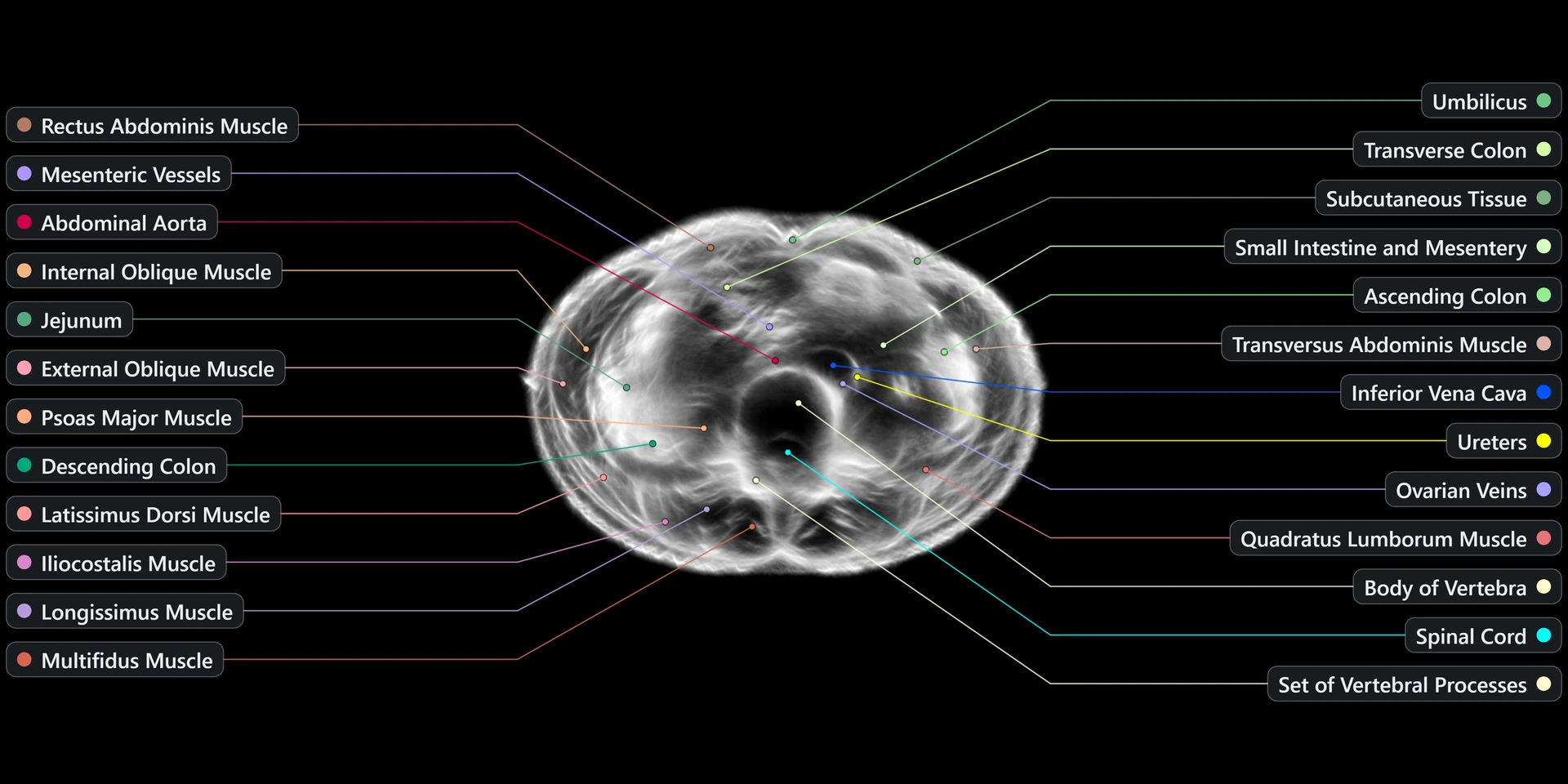
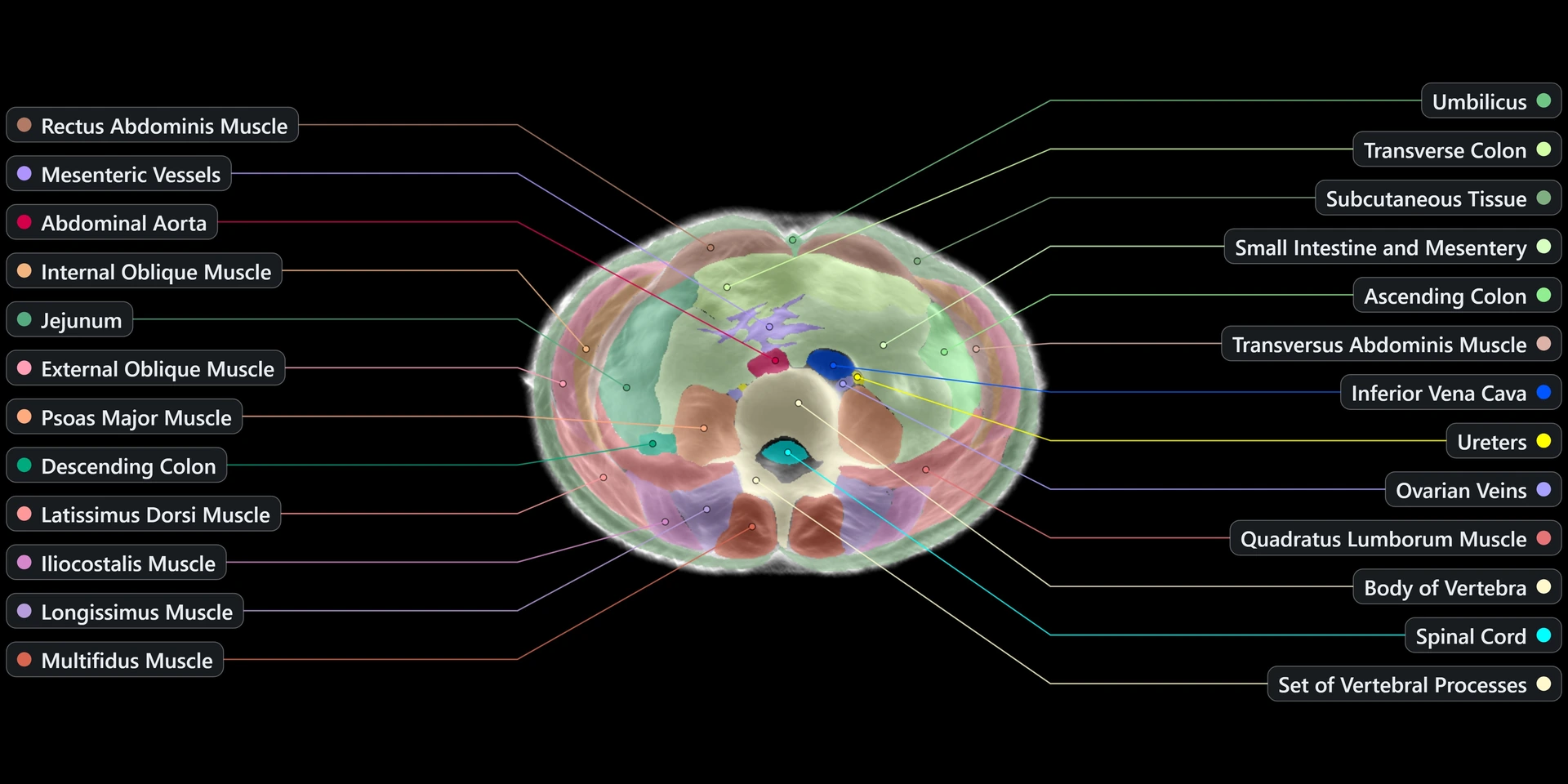
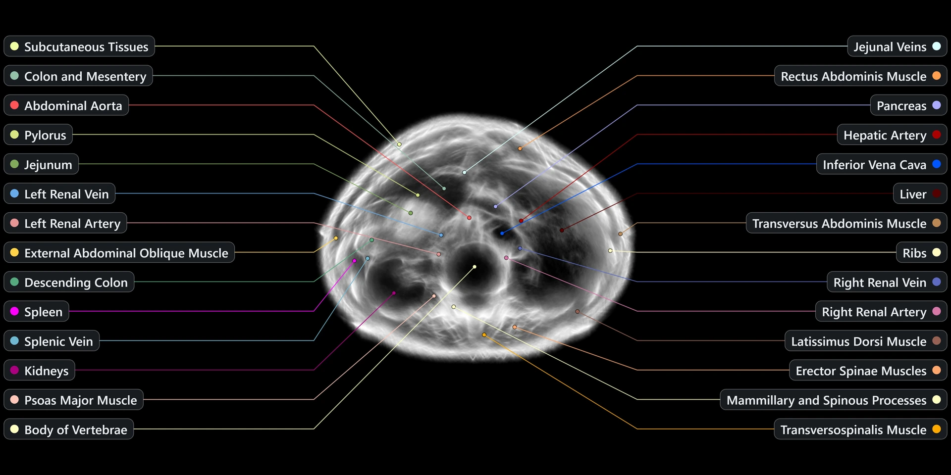
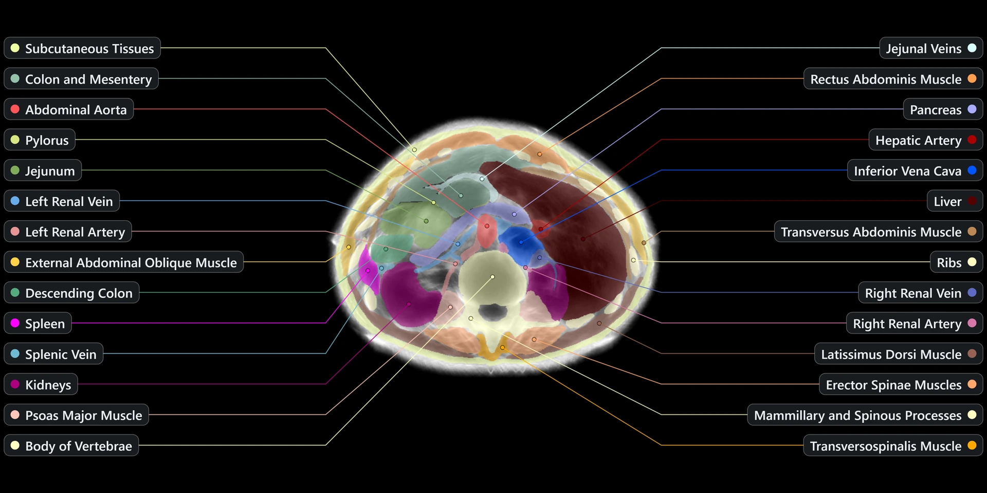
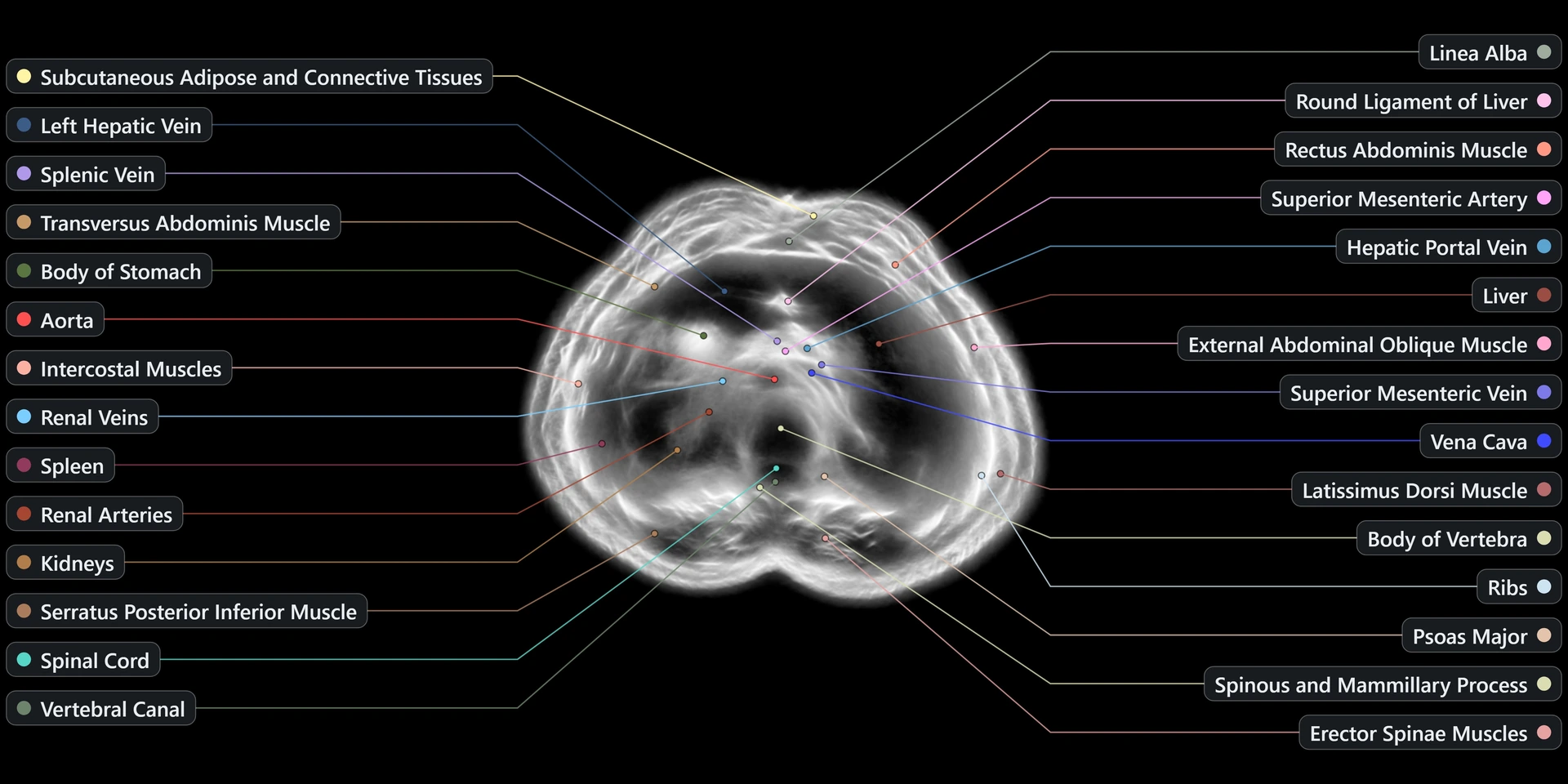
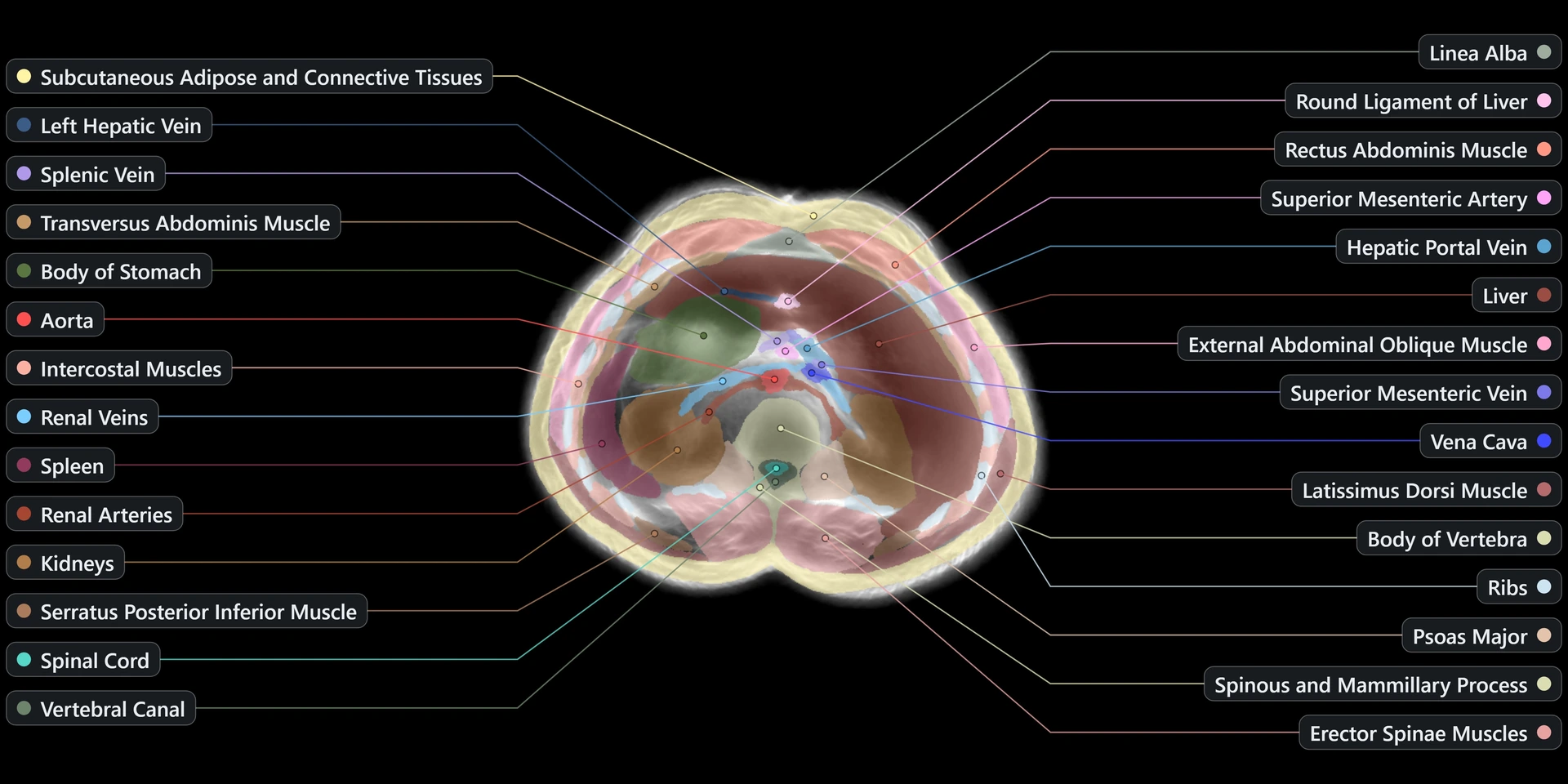
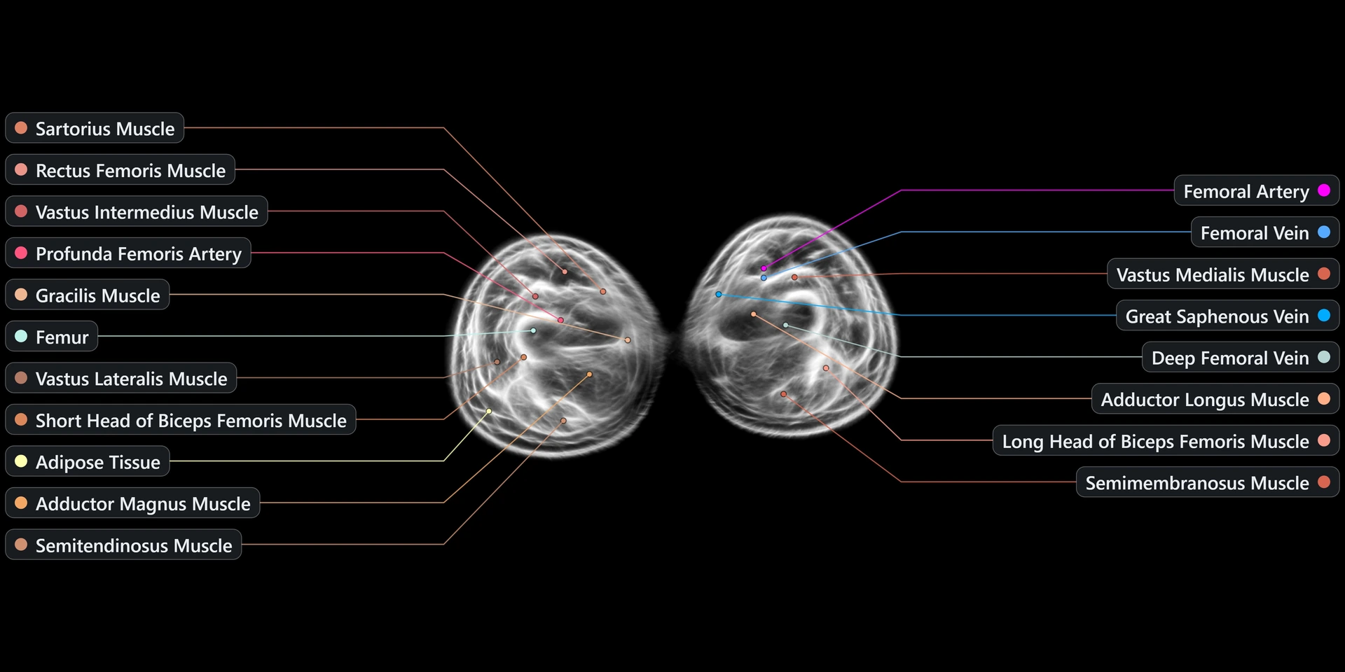
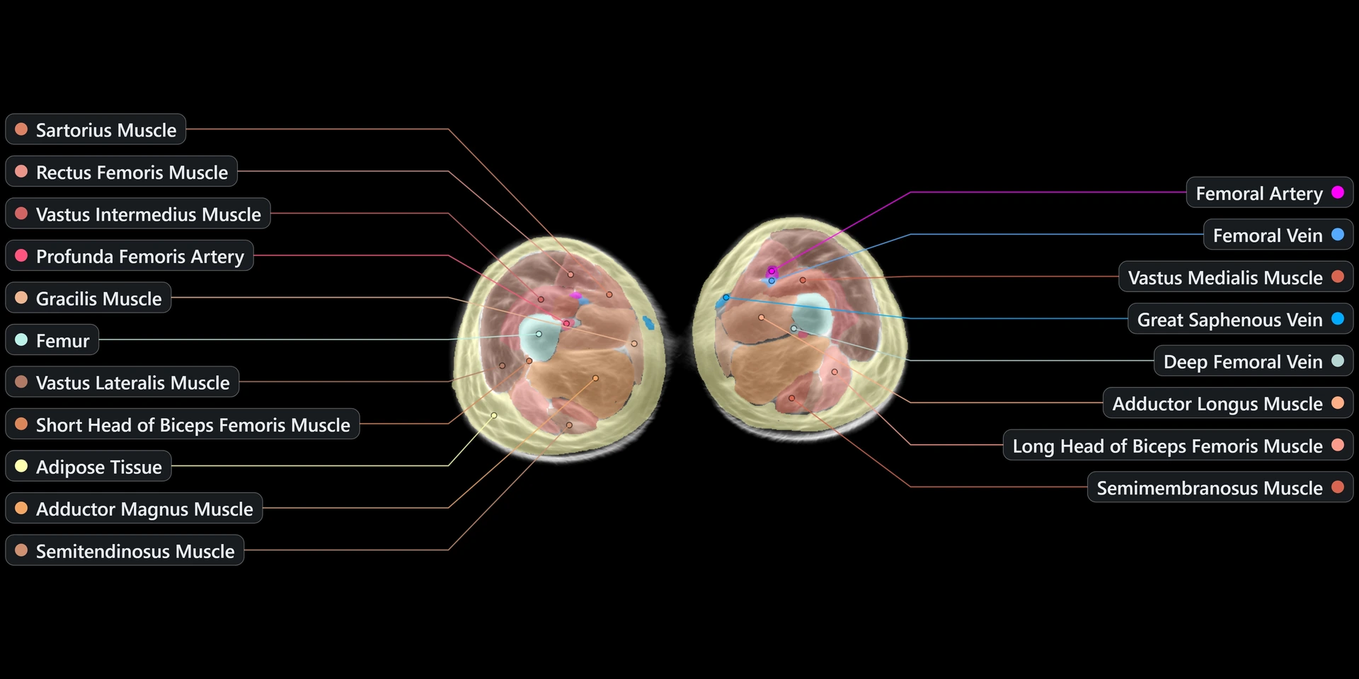
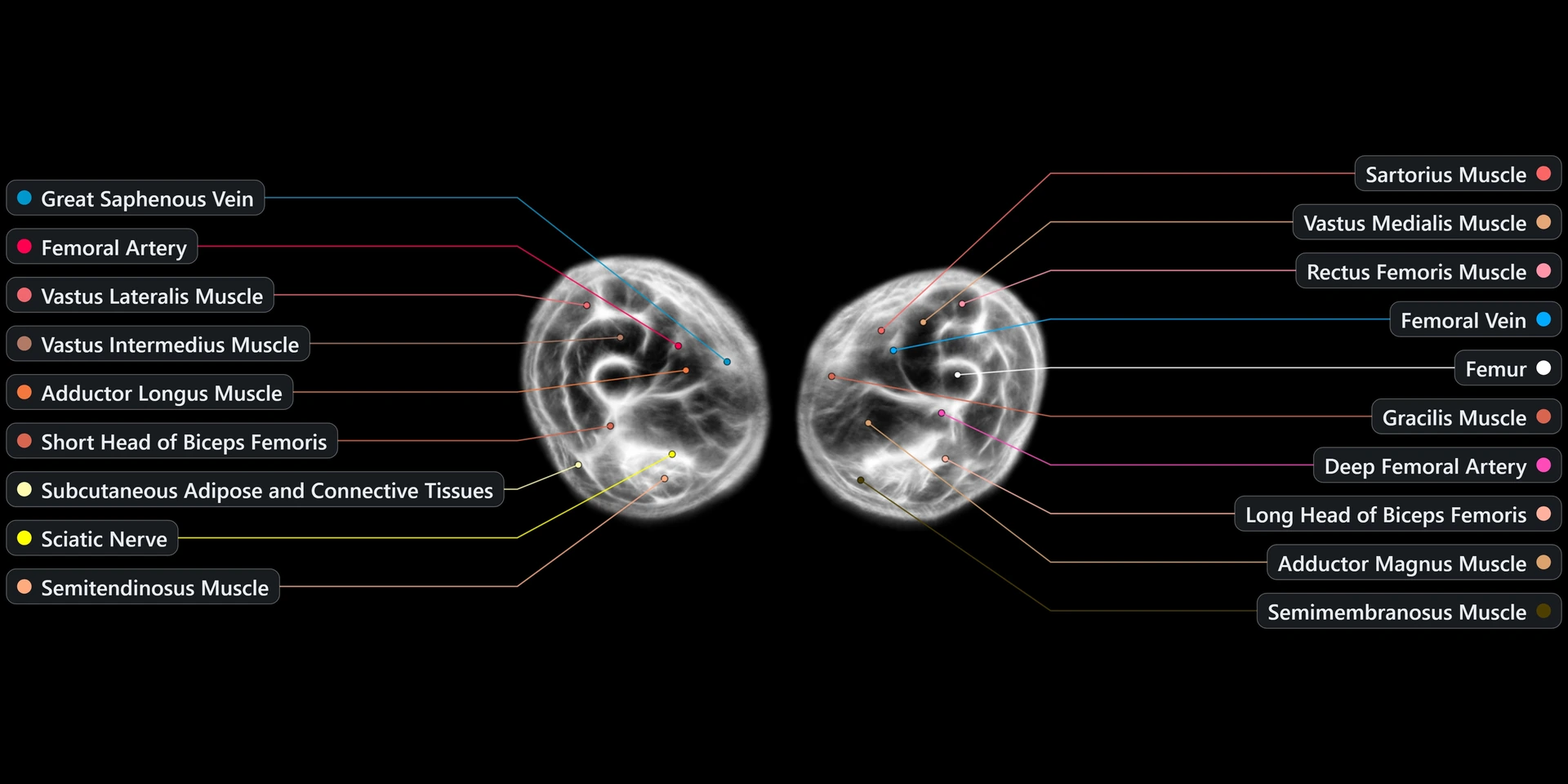
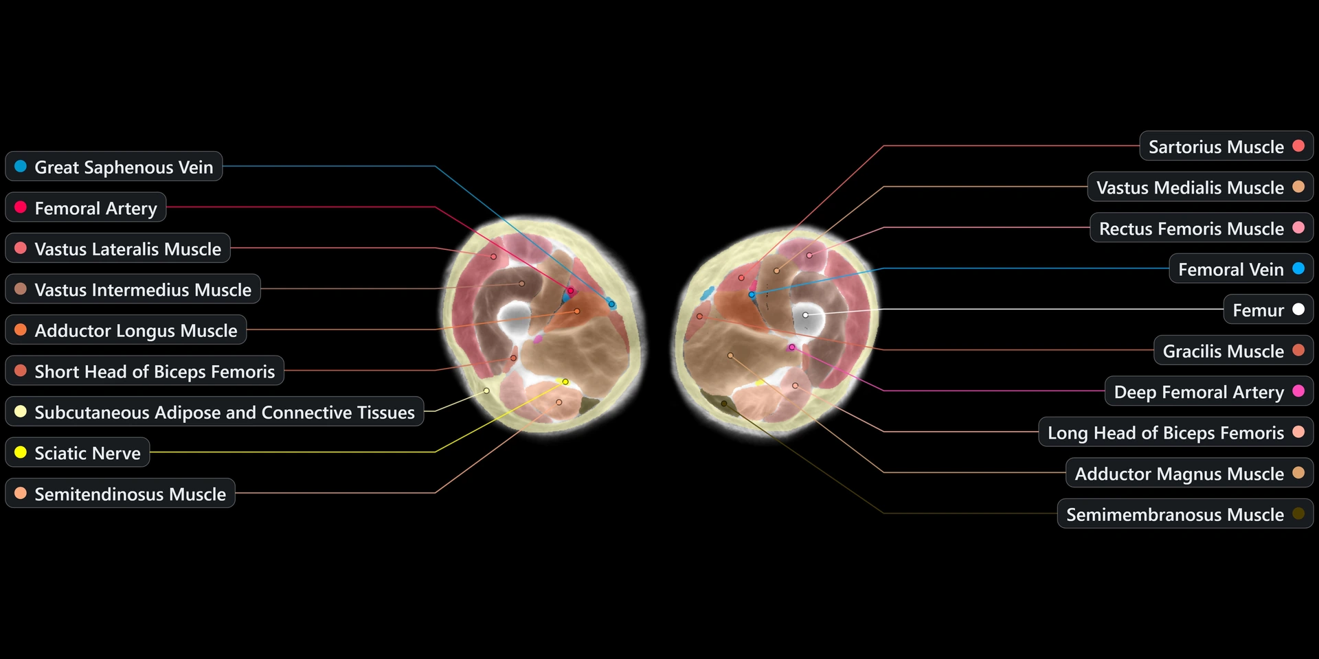
Five reconstructed slices, each crossfading between the raw scan and its labeled segmentation.
Swept body volumes
Reconstructed volumes played back slice by slice — the torso and the legs, each looping continuously.
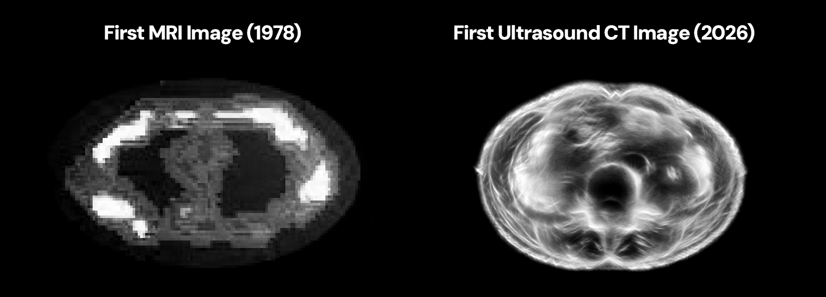
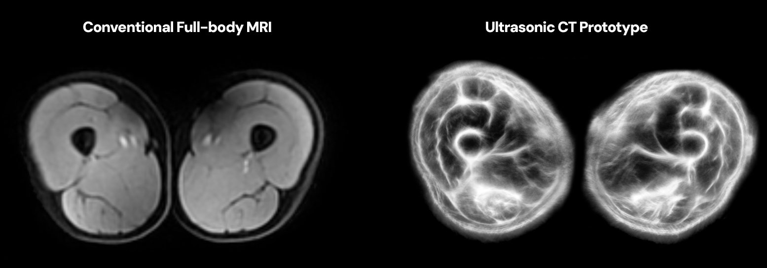
USCT vs MRI
Side-by-side comparisons of ultrasound-computed-tomography reconstructions against conventional MRI.
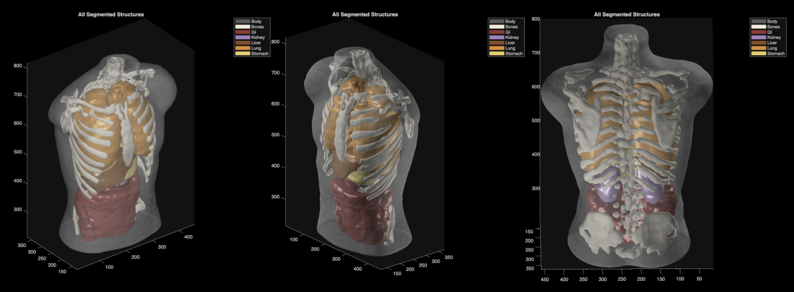
Phantom segmentation
A scan of an imaging phantom, segmented to validate how cleanly structures separate under controlled conditions.
Phantom sweeps — coronal & sagittal
Two sweeps through the same phantom volume, side by side: the coronal pass on the left and the sagittal pass on the right, looping together.